

Brain tumors, aneurysms, and other serious conditions in the brain often require immediate attention, including brain surgery. When you need a complex procedure like a craniotomy, you can trust the experts at WMCHealth.
Our cranial neurosurgeons have the experience and technology to support you through even the most challenging circumstances. Our surgical teams in New York are pioneers of advanced technologies and regularly perform brain surgery using minimally invasive techniques.
What is a Craniotomy?
A craniotomy is a surgical procedure that temporarily removes a section of the skull so a neurosurgeon can access the brain. This section, called a bone flap, is replaced and secured after treatment.
A craniotomy may be used to treat several conditions, including:
- Brain tumors
- Blood vessel abnormalities
- Infections
- Epilepsy
For brain tumors, surgery can help provide a diagnosis, relieve pressure, or remove as much tumor as possible. Your surgeon will explain the goals of your operation and whether additional treatment may be needed.
What to Know About Your Craniotomy
Your preparation, surgical approach, and recovery depend on your condition and the planned procedure.
How to Prepare for a Craniotomy
Our team reviews your diagnosis, imaging, and overall health before surgery. Specialized scans can help map out nearby areas involved in movement, language, vision, and other functions. Your surgical team will provide personalized instructions to help you prepare for your operation.
What to Expect During the Craniotomy
- You usually receive general anesthesia. You may be awake during portions of surgery for brain mapping and to test things like reflexes.
- The neurosurgeon makes an incision and temporarily removes a section of skull. The opening’s size and location depend on the condition being treated.
- The surgeon treats the affected area, using imaging, navigation, or mapping techniques when appropriate.
- After treatment, the bone flap is generally replaced and secured, and the incision is closed.
What to Expect After a Craniotomy
Your care team closely monitors you after surgery, sometimes initially in a neurological intensive care or step-down setting if needed. Imaging may be used to evaluate the results of surgery. Hospital stay and overall recovery vary with your procedure, health, and symptoms present before surgery.
- Fatigue and headache can occur during the early stages of recovery. Your team will explain expected symptoms and when it is appropriate to seek medical attention.
- You receive instructions about site care, medications, driving, exercise, and returning to work.
- Follow-up appointments and imaging help your team assess your recovery and plan any additional care as needed.
Advanced Techniques for Brain Tumor Surgery
WMCHealth offers a range of advanced technologies and techniques that may be used during a craniotomy when appropriate.
Brain Mapping and Awake Craniotomy
Brain mapping helps surgeons locate areas that control speech, movement, and other critical functions. Some patients remain awake during parts of surgery so the team can test these functions.
WMCHealth also uses AI-assisted mapping to show important connections within the brain. This information helps surgeons plan tumor removal and reduce the risk of injury.
Fluorescence-Guided Surgery
For certain brain tumors, a fluid called 5-ALA makes tumor tissue glow under a special light. This helps surgeons differentiate tumor tissue from nearby brain tissue, allowing the team to preserve as much healthy tissue as possible.
GammaTile Brachytherapy
GammaTile delivers targeted radiation through small tiles placed where a brain tumor was removed. The tiles gradually dissolve while radiation treats nearby tissue where tumor cells may remain. This approach limits radiation exposure to healthy brain tissue.
Laser Interstitial Thermal Therapy (LITT)
LITT uses a thin, MRI-guided laser probe to heat and destroy targeted brain tumor tissue. With this technique, you may be able to return home the next day, depending on your condition and recovery.
Risks of Craniotomy
As with any major operation, craniotomy has potential risks. These vary depending on the location and type of condition being treated. Craniotomy risks include:
- Bleeding or infection
- Seizures
- Leakage of cerebrospinal fluid, which surrounds the brain and spinal cord
- Blood clots or stroke
- Neurological changes, which may affect movement, speech, memory, or other functions
Your neurosurgeon will discuss the expected benefits and individual risks of your procedure. This discussion can help you understand your options and ask questions before surgery.

Vishad Sukul
Neurological Surgery
Locations:
-
100 Woods Rd ACP 4th Floor
Valhalla, NY 10595
 WMCHealth Provider
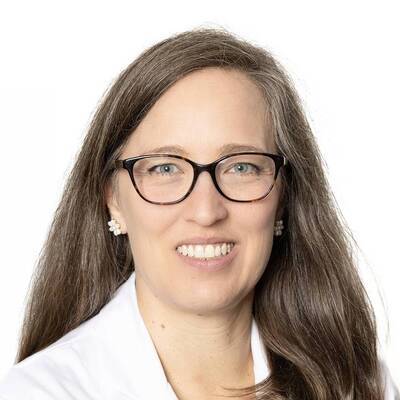
Katharine McNeill
Neuro-Oncology
Locations:
-
19 Bradhurst Avenue Suite 3850 S
Hawthorne, NY 10532
WMCHealth Provider
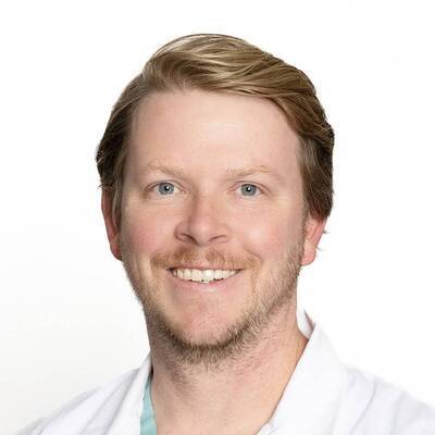
Davis Taylor
Neurological Surgery
Locations:
-
257 Lafayette Ave MOB Suite 285
Suffern, NY 10901
WMCHealth Provider

Justin Santarelli
Neuroendovascular Surgery, Neurological Surgery, Open Cerebrovascular Surgery
Locations:
-
100 Woods Rd ACP 4th Floor
Valhalla, NY 10595
WMCHealth Provider

Kalimullah Jan
Neurology, Neuroendovascular Surgery
Locations:
-
257 Lafayette Ave MOB Suite 285
Suffern, NY 10901
WMCHealth Provider

Tamara Benoit
Neurological Surgery
Locations:
-
100 Woods Rd ACP 4th Floor
Valhalla, NY 10595
WMCHealth Provider

Kelsey Kimble
Neurological Surgery
Locations:
-
100 Woods Rd ACP 4th Floor
Valhalla, NY 10595
WMCHealth Provider

Chirag Gandhi
Neuroendovascular Surgery, Neurological Surgery
Locations:
-
100 Woods Rd ACP 4th Floor
Valhalla, NY 10595
WMCHealth Provider
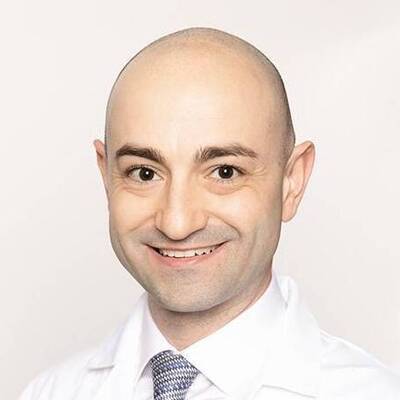
Sebastian Rubino
Neurological Surgery
Locations:
-
257 Lafayette Ave MOB Suite 285
Suffern, NY 10901
WMCHealth Provider
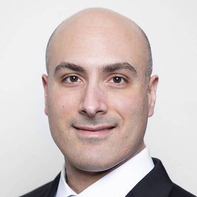
Merritt Kinon
Spine Surgery, Neurological Surgery
Locations:
-
100 Woods Rd ACP 4th Floor
Valhalla, NY 10595
WMCHealth Provider

Simon Hanft
Neurological Surgery
Locations:
-
100 Woods Rd ACP 4th Floor
Valhalla, NY 10595
WMCHealth Provider

Sherine Varghese
Neurological Surgery
Locations:
-
100 Woods Rd ACP 4th Floor
Valhalla, NY 10595
WMCHealth Provider

Stephanie Gandelman
Neurology, Neuroimmunology, Tele-Neurology
Locations:
-
19 Bradhurst Avenue Suite 3850 S
Hawthorne, NY 10532
WMCHealth Provider

Tatyana Gitlevich
Clinical Neurophysiology, Neurology, Epilepsy, Tele-Neurology
Locations:
-
19 Bradhurst Avenue Suite 3850 S
Hawthorne, NY 10532
WMCHealth Provider

Avinash Mohan
Neurological Surgery, Pediatric Neurosurgery, Pediatric Spine Surgery
Locations:
-
100 Crystal Run Road Suite 108
Middletown, NY 10941
WMCHealth Provider

Carrie Muh
Pediatric Neurosurgery
Locations:
-
19 Bradhurst Avenue Suite 3050
Hawthorne, NY 10532
WMCHealth Provider
Rachana Tyagi
Pediatric Spine Surgery, Neurological Surgery
Locations:
-
19 Baker Avenue Suite 302
Poughkeepsie, NY 12601
WMCHealth Provider
John Wainwright
Spine Surgery, Neurological Surgery
Locations:
-
100 Woods Rd ACP 4th Floor
Valhalla, NY 10595
WMCHealth Provider

Feliks Koyfman
Neurology, Vascular Neurology, Tele-Neurology
Locations:
-
19 Bradhurst Avenue Suite 3850 S
Hawthorne, NY 10532
WMCHealth Provider

Jared Pisapia
Pediatric Neurosurgery, Neurological Surgery
Locations:
-
100 Woods Rd ACP 4th Floor
Valhalla, NY 10595
WMCHealth Provider
Why Choose WMCHealth for Craniotomy
Our team brings personalized planning, advanced surgical tools, and collaboration to your care.
Individualized Surgical Planning
Your surgeon evaluates your condition and its relationship to important brain structures. The recommended approach considers your treatment goals, neurological function, and quality of life.
Techniques That Support Neurological Function
Imaging, computer-assisted navigation, and brain mapping help guide planning and surgery. Certain tumor operations may also use fluorescence guidance to help distinguish tumor tissue.
Collaboration Across Specialties
Neurosurgeons work with specialists in imaging, pathology, neurocritical care, and other fields to meet your exact needs. For brain tumors, care may also involve neuro-oncology and radiation oncology.